



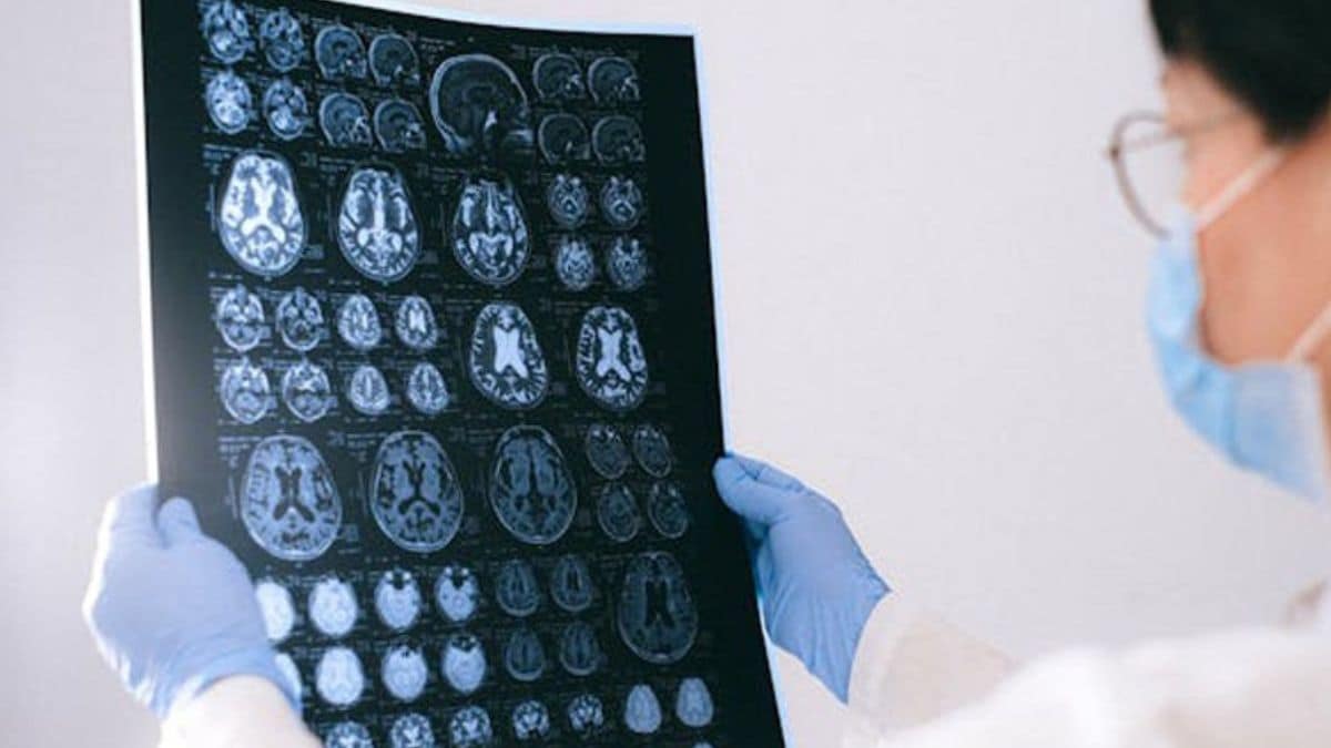
What began as a sudden weakness on the left side of her body initially looked like a classic stroke. But after two weeks of treatment yielded no relief and her condition steadily worsened, a 61-year-old woman from the Madurai–Virudhunagar region faced an alarming turn of events: specialised scans revealed a brain tumour. Seeking advanced surgical care, she travelled to Manipal Hospital Electronics City, where neurosurgeons discovered the mass lodged deep within her brain’s sensory cortex—a delicate region responsible for governing touch, spatial awareness, and bodily sensation.
In brain tumour surgery, precision is not simply about removing what can be seen. It is about knowing where the tumour ends and healthy, functioning brain begins. This is particularly challenging when a tumour lies close to areas that control essential functions such as movement and sensation. Such constant need for greater accuracy has driven the development of newer surgical adjuncts that can give neurosurgeons information beyond what the naked eye can provide. One such technology is 5-aminolevulinic acid (5-ALA) fluorescence-guided surgery, which is helping surgeons better visualise tumour tissue during an operation.
“Given the critical location, the surgical team used multiple adjuncts, neuronavigation, 5-ALA fluorescence-guided surgery and neuromonitoring, to guide the procedure. These technologies helped the team remove the majority of the tumour while navigating the surrounding functional brain tissue. The patient had a favourable molecular analysis and is now planned for chemotherapy and radiotherapy,” explains Dr Shyam Duvuru, Senior Consultant - Neurosurgery, Manipal Hospital Electronics City.
5-ALA is a compound that, when administered before surgery, is preferentially taken up by certain brain tumour cells. When viewed under a specialised blue light fluorescence in the Carl Zeiss KINEVO 900 operating microscope, these tumour cells fluoresce, standing out clearly from surrounding normal brain tissue. This advanced visualisation serves as an important intraoperative aid during surgery, particularly at the tumour margins, supporting the surgical team in their efforts to identify and remove tumour tissue as precisely as possible with greater precision.
For the patient, the goal is to achieve as complete a resection as safely possible, while preserving critical brain functions. The value of such adjuncts becomes especially important when tumours are located in eloquent areas of the brain, regions responsible for vital functions. In these situations, surgery involves a delicate balance between removing as much tumour as possible and protecting the surrounding healthy tissue.
The evolution of brain tumour surgery is increasingly about combining surgical expertise with technologies that provide the surgeon with better information at critical moments. 5-ALA fluorescence is one such advance, turning what may be difficult to distinguish by sight alone into an additional visual guide inside the operating room.
Notably, this procedure marked a significant milestone as the first reported use of 5-ALA fluorescence-guided surgery in Karnataka and South India, performed using the Carl Zeiss KINEVO 900 surgical microscope, further advancing the adoption of fluorescence-guided technology in brain tumour surgery.
For patients facing complex brain tumours, these advances represent an important step towards more informed, precise and safer surgery, where every available tool can contribute to the best possible outcome.