Researchers in the US have created the first detailed map of the changes at the molecular level in cells that lead to embryonic development.
The research, published in the journal Science, used the latest technology from the emerging field of single cell biology -- the study of all chemical processes happening at the cellular level -- to profile more than 80,000 cells in the embryo of the worm Caenorhabditis elegans.
"Over the past few years, new single cell genomics methods have revolutionised the study of animal development," said John I Murray from the University of Pennsylvania in the US.
"Our study takes advantage of the fact that the C. elegans embryo has a very small number of cells produced by a known and completely reproducible pattern of cell divisions. Using single cell genomics methods, we were able to identify over 87 per cent of embryonic cells from gastrulation (when there are about 50 cells present) through the end of embryogenesis," he added.
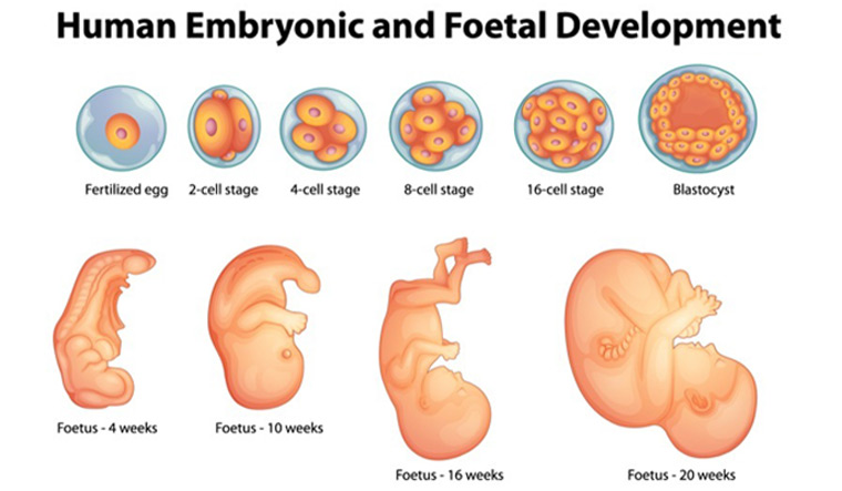
The worm hatches with only 558 cells in its body, and like every multicellular organism, each of its cells is derived from division from a single fertilized egg.
From this, a "cell lineage tree" can be made, showing the replication history of every cell, along with their relationship to each other, the study mentioned.
The researchers also characterised the molecular chemistry at the level of individual cells by mapping all the RNAs in a cell during development -- using the single cell genomics approach.


