I’m seeing double,” Rita said, sitting across from me. Her eyes were doing a passable impression of a Picasso painting. She was dressed in a white dress with silver streaks, and a grey hairband kept her highlighted fringe off her forehead—one less thing to be seeing double of.
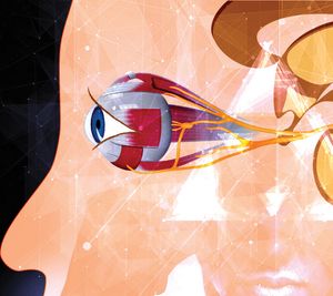
“It is unusual for someone in their 30s to have double vision,” I stated. “It’s only when I try and look in a certain direction,” she clarified. I examined her to find that her eyes were slightly misaligned, the right eye being ‘down and out’—a classic sign of the oculomotor nerve being affected. This is the third among the 12 pairs of cranial nerves. It is also one of the three nerves responsible for moving the eyes. “Oftentimes, diabetes is a cause, but we have to make sure nothing is pressing against the nerve from outside,” I explained to her and her father, who by now was visibly concerned when I pointed out the subtle drooping of the eyelid.
A few days later, they returned with the MRI and CT scans I’d ordered, and as suspected, it showed an aneurysm pressing against the nerve. An aneurysm is an abnormal dilation of an artery due to its wall being weak, causing it to balloon out. “This is what’s causing your problem,” I said, pointing to the one centimetre blob. “Given its anatomy and configuration, it is best treated by surgery, or else I would have suggested an endovascular route to try and fix it by inserting a flow diverter,” I explained, adding that the risk of rupture is high and that it could sometimes be fatal. After a round of detailed questioning and a couple of second opinions later, they agreed to operate.
On a cool winter morning, we wheeled her into the operation theatre. The sterile air inside the room seeming colder than usual, which can sometimes happen when one is nervous before a tough case. Aneurysms are always roiled with a tempestuous secret. To identify them, to dissect around them, and then to finally clip them can either be a tranquil sea or a tsunami.
After fixing the head on a clamp, we opened the skull in the usual fashion, the lightly pulsating frontal and temporal lobes lying exposed in front of me as I gently separated the delicate veil of silver strands holding the two lobes together, my instruments gleaming like silver serpents dancing against the brilliant light of the microscope. The oculomotor nerve had been pushed into a corner by the throbbing crimson ball that appeared more malevolent in reality than on a black and white scan, its fragile pulsing wall threatening to rupture at any point and shatter the icy silence of the room. I gained control of the main artery from which it arose and placed a temporary clip on it like a dam, reducing the pressure of blood flowing into the aneurysm, so that I could dissect around it with impunity. “That’s the neck we need to throttle,” I told my assistant, channelling my inner gladiator. A 7mm minuscule clip, made from the most resilient titanium, was brought into play.
With steady hands, I placed the prongs precisely across the entire length of the neck, and like the talons of an eagle, closed it shut. A subtle shift, a barely perceptible tightening, and the bulging wall ceased its ominous throbbing. The reddish bloom, once a beacon of impending doom, paled, its dangerous pulse silenced. The flow of life, diverted from its perilous detour, resumed its proper course. The oculomotor nerve was relieved of the burden it was bearing.
Rita made a remarkable recovery and was discharged in a few days. When she returned three months later, she gave me a piercing look, her eyes no longer seeing double. The eyelids were in perfect symmetry. The third nerve was working in sync with the fourth, fifth, and sixth.
The author is consultant neurosurgeon at Wockhardt Hospital, Mumbai.
mazdaturel@gmail.com @mazdaturel


